Identify the structure in the diagram below
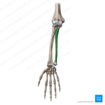
The Correct Answer is ["Ulna bone"]
The ulna is one of the two bones of the forearm, located on the medial (inner) side of the arm.
It is a long bone that runs parallel to the radius bone, extending from the elbow joint to the wrist joint.
The ulna features several surface landmarks, including the olecranon process, which forms the bony tip of the elbow.
The bone is also involved in the formation of the elbow joint, where it articulates with the humerus bone, and the wrist joint, where it articulates with the radius bone and several carpal bones.
The ulna is an important site for the attachment of muscles involved in forearm and wrist movements.
Nursing Test Bank
Naxlex Comprehensive Predictor Exams
Related Questions
Correct Answer is C
Explanation
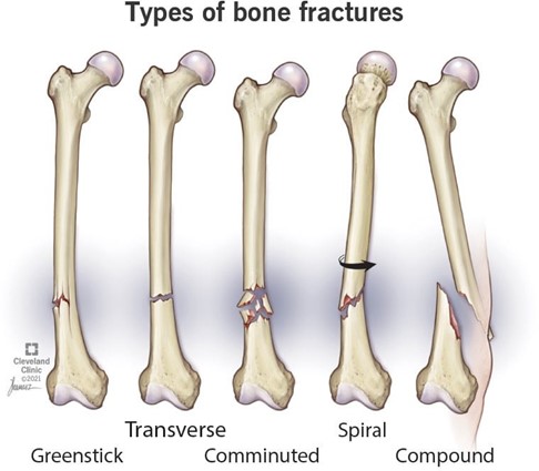
Choice A is incorrect because transverse fractures are the type of fractures where the bone has a horizontal fracture line.
They are usually caused by a strong force applied at a right angle to the bone shaft.
Choice B is incorrect because spiral fractures are the type of fractures where the bone has an angled fracture line that curves around the bone shaft.
They are usually caused by a twisting force applied to the bone.
Choice D is incorrect because greenstick fractures are the type of fractures where the bone is bent and partially broken on one side.
They are usually seen in children whose bones are softer and more flexible than adults.
Correct Answer is ["Occipital bone"]
Explanation

The occipital bone is a flat, unpaired bone located at the posterior aspect of the skull, forming the lower part of the back of the head.
It features several surface landmarks, including the external occipital protuberance, which serves as an attachment site for muscles and ligaments.
The occipital bone also contains several foramina, including the foramen magnum, which allows the spinal cord to pass through and connect to the brain.
The occipital bone is an important site for the attachment of muscles involved in head movement and posture.
Whether you are a student looking to ace your exams or a practicing nurse seeking to enhance your expertise , our nursing education contents will empower you with the confidence and competence to make a difference in the lives of patients and become a respected leader in the healthcare field.
Visit Naxlex, invest in your future and unlock endless possibilities with our unparalleled nursing education contents today