Identify the anatomical movement illustrated below and state the joint involved (for example flexion of the knee joint).
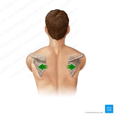
The Correct Answer is ["Scapula protraction at the scapulothoracic joint."]
Scapula protraction refers to the movement of the shoulder blade (scapula) away from the spine, toward the front of the body.
This movement is also known as scapular abduction or anterior scapular tilt.
The joint involved in scapula protraction is the scapulothoracic joint, which is not a true joint, but rather a functional joint formed by the articulation between the scapula and the thorax.
The scapula is a flat bone that glides over the back of the ribcage, allowing for a wide range of movements of the arm.
Other movements of the scapulothoracic joint include:
Scapula retraction: This refers to the movement of the shoulder blade towards the spine, away from the front of the body.
This movement is also known as scapular adduction or posterior scapular tilt.
Scapula elevation: This refers to the movement of the shoulder blade upwards towards the ears.
This movement is also known as the upward rotation of the scapula.
Scapula depression: This refers to the movement of the shoulder blade downwards towards the feet.
This movement is also known as a downward rotation of the scapula.
Scapula upward tilt: This refers to the movement of the upper border of the shoulder blade upwards, towards the head.
This movement is also known as superior scapular rotation.
Scapula downward tilt: This refers to the movement of the upper border of the shoulder blade downwards, towards the feet.
This movement is also known as inferior scapular rotation.
All of these movements are important for proper shoulder function and are necessary for a wide range of daily activities, such as reaching, lifting, pushing, and pulling.
Nursing Test Bank
Naxlex Comprehensive Predictor Exams
Related Questions
Correct Answer is C
Explanation
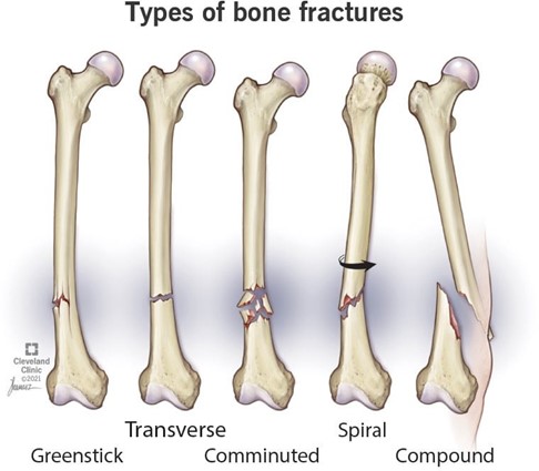
Choice A is incorrect because transverse fractures are the type of fractures where the bone has a horizontal fracture line.
They are usually caused by a strong force applied at a right angle to the bone shaft.
Choice B is incorrect because spiral fractures are the type of fractures where the bone has an angled fracture line that curves around the bone shaft.
They are usually caused by a twisting force applied to the bone.
Choice D is incorrect because greenstick fractures are the type of fractures where the bone is bent and partially broken on one side.
They are usually seen in children whose bones are softer and more flexible than adults.
Correct Answer is ["Mandible protraction of the temporomandibular joint (TMJ)."]
Explanation
Mandible protraction refers to the movement of the lower jaw (mandible) forwards, away from its rest position.
This movement is also known as jaw thrust.
The joint involved in mandible protraction is the temporomandibular joint (TMJ), which is a synovial joint that connects the mandible to the temporal bone of the skull.
This joint allows for a variety of movements, including:
Mandible retraction: This refers to the movement of the lower jaw backward, towards the skull.
Mandible elevation: This refers to the movement of the lower jaw upwards, towards the upper jaw.
Mandible depression: This refers to the movement of the lower jaw downwards, away from the upper jaw.
Mandible lateral excursion: This refers to the movement of the lower jaw to either the left or the right.
Mandible medial excursion: This refers to the movement of the lower jaw back to its midline position after a lateral excursion.
These movements of the TMJ are essential for functions such as chewing, speaking, and swallowing.
However, excessive or repetitive movements of the jaw can lead to TMJ disorders, which can cause pain, clicking, popping, or locking of the jaw.
Whether you are a student looking to ace your exams or a practicing nurse seeking to enhance your expertise , our nursing education contents will empower you with the confidence and competence to make a difference in the lives of patients and become a respected leader in the healthcare field.
Visit Naxlex, invest in your future and unlock endless possibilities with our unparalleled nursing education contents today