Assessment of a pregnant woman reveals a pigmented vertical line between the umbilicus and the pubis. The nurse documents this finding as:
Select one:
Vascular spider veins.
Linea nigra.
Melasma.
Striae gravidarum.
The Correct Answer is B
Choice A Reason: Vascular spider veins. This is an incorrect answer that refers to a different skin change that occurs during pregnancy, which affects the blood vessels, not the pigment. Vascular spider veins are small red or purple clusters of blood vessels that appear on the skin, especially on the face, neck, chest, or legs. Vascular spider veins are caused by increased blood volume and hormonal changes, which dilate and rupture the capillaries. Vascular spider veins are harmless and usually disappear after delivery.
Choice B Reason: Linea nigra. This is because linea nigra is a term that refers to a darkened vertical line that appears on the abdomen during pregnancy, which runs from the umbilicus to the pubis. Linea nigra is caused by increased production of melanin, which is a pigment that gives color to the skin and hair. Linea nigra is more common and noticeable in women with darker skin tones, and it usually fades after delivery.
Choice C Reason: Melasma. This is an incorrect answer that refers to a different skin change that occurs during pregnancy, which affects the pigment, but not in a linear patern. Melasma is a term that refers to patches of brown or gray-brown discoloration that appear on the face, especially on the forehead, cheeks, nose, or upper lip. Melasma is also caused by increased production of melanin, but it is influenced by sun exposure and genetic factors. Melasma is also known as chloasma or the mask of pregnancy, and it may persist after delivery.
Choice D Reason: Striae gravidarum. This is an incorrect answer that refers to a different skin change that occurs during pregnancy, which affects the connective tissue, not the pigment. Striae gravidarum are stretch marks that appear on the skin, especially on the abdomen, breasts, hips, or thighs. Striae gravidarum are caused by rapid growth and stretching of the skin, which damage the collagen and elastin fibers. Striae gravidarum are initially red or purple, but they fade to white or silver after delivery.

Nursing Test Bank
Naxlex Comprehensive Predictor Exams
Related Questions
Correct Answer is C
Explanation
Choice A Reason: Manifestations of uteroplacental insufficiency. This is an incorrect answer that describes a different condition that affects the fetus, not the mother. Uteroplacental insufficiency is a condition where the placenta fails to deliver adequate oxygen and nutrients to the fetus, which can result in fetal growth restriction, distress, or demise. Uteroplacental insufficiency does not cause shortness of breath, hypoxia, or cyanosis in the mother.
Choice B Reason: Manifestations of prolapsed cord. This is an incorrect answer that refers to another condition that affects the fetus, not the mother. Prolapsed cord is a condition where the umbilical cord slips through the cervix before the baby and becomes compressed by the fetal head, which can reduce oxygen flow to the fetus. Prolapsed cord does not cause shortness of breath, hypoxia, or cyanosis in the mother.
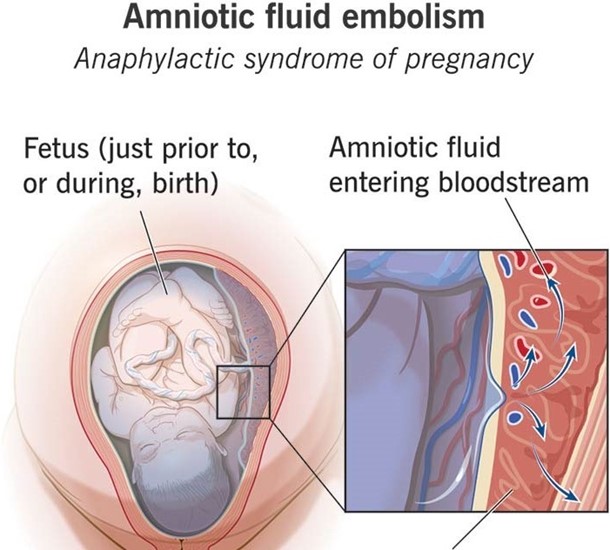
Choice C Reason: Manifestations of anaphylactoid syndrome of pregnancy. This is because anaphylactoid syndrome of pregnancy, also known as amniotic fluid embolism, is a rare and fatal condition where amniotic fluid enters into the maternal bloodstream and causes an allergic reaction, which can lead to respiratory failure, cardiac arrest, coagulopathy, and coma. Anaphylactoid syndrome of pregnancy can occur during or after labor and delivery, especially in cases of NSVD, multiparity, advanced maternal age, or placental abruption.
Choice D Reason: Manifestations of an acute asthmatic episode. This is an incorrect answer that assumes that the mother has a history of asthma or an allergic trigger. Asthma is a chronic inflammatory disorder of the airways that causes wheezing, coughing, chest tightness, and dyspnea. Asthma can be exacerbated by pregnancy or labor, but it is not a common cause of sudden onset respiratory distress in the postpartum period.

Correct Answer is A
Explanation
Choice A Reason: Two arteries, one vein. This is because two arteries and one vein are the normal components of the umbilical cord, which is a structure that connects the fetus to the placenta and provides blood circulation between them. The umbilical cord carries oxygenated blood from the placenta to the fetus through the umbilical vein, and deoxygenated blood from the fetus to the placenta through the umbilical arteries.
Choice B Reason: Two veins, one artery. This is an incorrect answer that indicates an abnormal anatomy of the umbilical cord, which is known as single umbilical artery (SUA). SUA is a condition where there is only one umbilical artery instead of two, which can reduce blood flow and oxygen delivery to the fetus. SUA can be associated with congenital anomalies or growth restriction in some cases.
Choice C Reason: Two veins, two arteries. This is an incorrect answer that indicates an abnormal anatomy of the umbilical cord, which is known as double umbilical vein (DUV). DUV is a condition where there are two umbilical veins instead of one, which can increase blood flow and oxygen delivery to the fetus. DUV can be associated with fetal overgrowth or polycythemia in some cases.
Choice D Reason: One artery, one vein. This is an incorrect answer that indicates an abnormal anatomy of the umbilical cord, which is also known as single umbilical artery (SUA). SUA is a condition where there is only one umbilical artery instead of two, which can reduce blood flow and oxygen delivery to the fetus. SUA can be associated with congenital anomalies or growth restriction in some cases.
Whether you are a student looking to ace your exams or a practicing nurse seeking to enhance your expertise , our nursing education contents will empower you with the confidence and competence to make a difference in the lives of patients and become a respected leader in the healthcare field.
Visit Naxlex, invest in your future and unlock endless possibilities with our unparalleled nursing education contents today
Report Wrong Answer on the Current Question
Do you disagree with the answer? If yes, what is your expected answer? Explain.
Kindly be descriptive with the issue you are facing.
