
A nurse is collecting data from an infant a large patent ductus arteriosus. Which of the following is clinical manifestations should the nurse expect?
Machine like murmur
Chronic hypoxemia
Cyanosis with crying
Weak pulse
The Correct Answer is A
A. Machine-like murmur.
This option is correct. A characteristic clinical manifestation of a large patent ductus arteriosus is a continuous "machine-like" murmur heard on auscultation. This murmur is typically heard best at the upper left sternal border and may radiate to the back.
B. Chronic hypoxemia.
Chronic hypoxemia is not typically a primary manifestation of a large PDA. While PDA can lead to increased pulmonary blood flow and potentially contribute to pulmonary congestion, chronic hypoxemia may not be a prominent feature unless complications such as heart failure develop.
C. Cyanosis with crying.
Cyanosis with crying is more commonly associated with cyanotic congenital heart defects such as tetralogy of Fallot. While PDA can contribute to cyanosis in certain circumstances, it is not typically a consistent clinical manifestation.
D. Weak pulse.
A weak pulse is not typically associated specifically with a large PDA. Infants with PDA may have bounding pulses due to increased blood flow through the ductus arteriosus.
Nursing Test Bank
Naxlex Comprehensive Predictor Exams
Related Questions
Correct Answer is C
Explanation
A. Machine-like murmur:
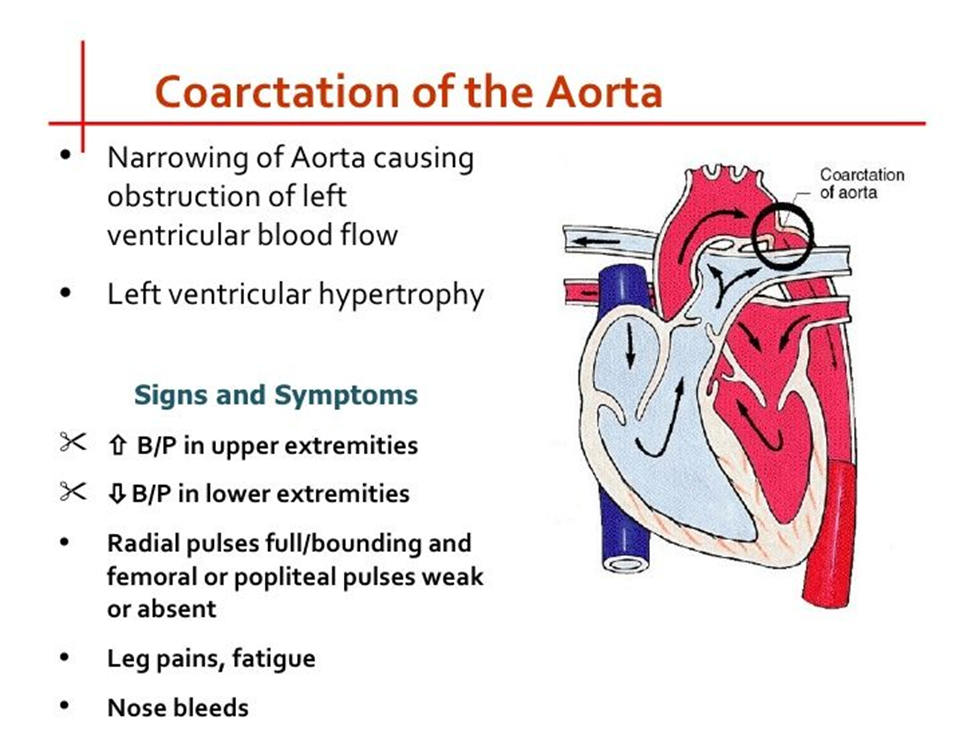
A machine-like murmur typically refers to a continuous murmur, which can be heard throughout systole and diastole. While machine-like murmurs can be associated with certain cardiac conditions, such as patent ductus arteriosus (PDA), they are not typically heard in coarctation of the aorta. In coarctation of the aorta, a systolic ejection murmur may be heard over the upper left sternal border due to turbulent blood flow across the narrowed aortic segment.
B. Severe cyanosis:
Cyanosis refers to a bluish discoloration of the skin and mucous membranes due to decreased oxygenation of the blood. While cyanosis can occur in various congenital heart defects, such as tetralogy of Fallot, it is not a characteristic manifestation of coarctation of the aorta. Coarctation of the aorta typically results in decreased blood flow to the lower extremities rather than mixing of oxygenated and deoxygenated blood.
C. Decreased blood pressure in the legs:
This is the correct choice. Coarctation of the aorta is characterized by narrowing of the aorta, which leads to decreased blood flow to the lower extremities. Consequently, blood pressure measurements in the legs may be lower compared to those in the arms. This finding is often a key indicator of coarctation of the aorta.
D. Pulmonary edema:
Pulmonary edema refers to the accumulation of fluid in the lungs and is typically associated with conditions such as heart failure or fluid overload. While some congenital heart defects may lead to heart failure and subsequent pulmonary edema, coarctation of the aorta does not directly cause pulmonary edema. Instead, it primarily affects blood flow to the lower extremities due to the narrowing of the aorta.
Correct Answer is C
Explanation
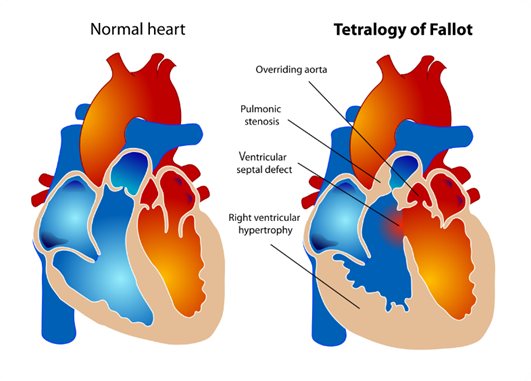
A. Overriding aorta: In Tetralogy of Fallot, the aorta is positioned over the ventricular septal defect (VSD), rather than solely over the left ventricle as it would be in a normal heart. This is called overriding aorta, which allows blood from both the right and left ventricles to enter the aorta.
B. Pulmonary stenosis: This is a critical component of Tetralogy of Fallot. Pulmonary stenosis refers to narrowing of the pulmonary valve or the area just below it, which restricts blood flow from the right ventricle to the pulmonary artery. This results in decreased blood flow to the lungs for oxygenation.
C. Left ventricular hypertrophy: This choice is not typically associated with Tetralogy of Fallot. Left ventricular hypertrophy refers to an enlargement or thickening of the muscular wall of the left ventricle of the heart. It is often seen in conditions where the left ventricle has to work harder to pump blood, such as in hypertension or aortic stenosis, but it is not a characteristic feature of Tetralogy of Fallot.
D. Ventricular septal defect: This defect is one of the four components of Tetralogy of Fallot. A ventricular septal defect (VSD) is a hole in the septum, the muscular wall that separates the left and right ventricles of the heart. In Tetralogy of Fallot, the VSD allows oxygen-poor blood from the right ventricle to flow directly into the left ventricle and out to the body.
Whether you are a student looking to ace your exams or a practicing nurse seeking to enhance your expertise , our nursing education contents will empower you with the confidence and competence to make a difference in the lives of patients and become a respected leader in the healthcare field.
Visit Naxlex, invest in your future and unlock endless possibilities with our unparalleled nursing education contents today