A client who recently suffered a stroke suffers from right-sided homonymous hemianopsia. What is the best action for the nurse to take when caring for the client during mealtime?
Place food trays on the left side of the client.
Place food trays on the right side of the client.
Perform a focused visual exam.
Have the assistive personnel feed all meals to the client.
The Correct Answer is A
Choice A reason: This is the correct answer because right-sided homonymous hemianopsia means that the client has lost vision in the right half of both eyes, so placing food trays on the left side of the client will help them see and access their food better.
Choice B reason: This is incorrect because placing food trays on the right side of the client will make it harder for them to see and reach their food, as they have no vision on that side.
Choice C reason: This is incorrect because performing a focused visual exam is not an appropriate action for the nurse to take during meal time. The nurse should assess the client's vision before or after meals, but not interfere with their eating.
Choice D reason: This is incorrect because having the assistive personnel feed all meals to the client will decrease their independence and dignity, as well as their ability to practice using their unaffected side. The nurse should encourage and assist the client to feed themselves as much as possible, and only provide assistance when needed.
Nursing Test Bank
Naxlex Comprehensive Predictor Exams
Related Questions
Correct Answer is B
Explanation
Choice A Reason: The test is not inconclusive, but rather positive for conductive hearing loss. The Weber test involves placing a vibrating tuning fork on the center of the forehead and asking the client which ear hears the sound louder. It can help differentiate between conductive and sensorineural hearing loss.
Choice B Reason: This is the correct choice. The client has conductive hearing loss, which is a type of hearing loss that occurs when sound waves are blocked or reduced in the outer or middle ear. It can be caused by earwax, infection, fluid, perforation, or trauma. In conductive hearing loss, the Weber test shows lateralization to the affected ear, meaning the sound is heard louder in that ear.
Choice C Reason: The client does not have normal hearing, but rather conductive hearing loss. In normal hearing, the Weber test shows no lateralization, meaning the sound is heard equally in both ears.
Choice D Reason: The client does not have sensorineural hearing loss, but rather conductive hearing loss. Sensorineural hearing loss is a type of hearing loss that occurs when there is damage to the inner ear or auditory nerve. It can be caused by aging, noise exposure, disease, or drugs. In sensorineural hearing loss, the Weber test shows lateralization to the unaffected ear, meaning the sound is heard louder in that ear.
Correct Answer is B
Explanation
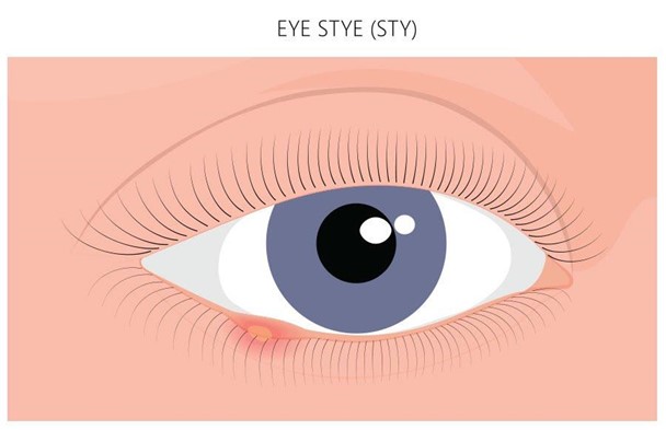
Choice A Reason: An antifungal cream is not indicated for a sty, which is an infection of the eyelash follicle or sebaceous gland caused by bacteria.
Choice B Reason: This is the correct answer because warm compresses can help relieve pain and inflammation, and promote drainage of the sty.
Choice C Reason: Ice and cold compresses are not recommended for a sty, as they can constrict blood vessels and delay healing.
Choice D Reason: There is no need to test the other eye for vision loss, as a sty does not affect vision unless it is very large or obstructs the pupil.

Whether you are a student looking to ace your exams or a practicing nurse seeking to enhance your expertise , our nursing education contents will empower you with the confidence and competence to make a difference in the lives of patients and become a respected leader in the healthcare field.
Visit Naxlex, invest in your future and unlock endless possibilities with our unparalleled nursing education contents today
Report Wrong Answer on the Current Question
Do you disagree with the answer? If yes, what is your expected answer? Explain.
Kindly be descriptive with the issue you are facing.
