Which muscle of the quadriceps femoris group lies on the side surface of the lower extremities?
Anterior tibialis
Rectus femoris
Gastrocnemius
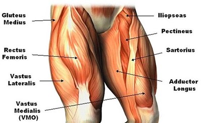
Vastus lateralis
The Correct Answer is D
The quadriceps femoris group is a group of four muscles located in the anterior compartment of the thigh. These muscles are responsible for extending the leg at the knee joint. The four muscles that make up the quadriceps femoris group are the rectus femoris, vastus medialis, vastus intermedius, and vastus lateralis.
The vastus lateralis is the largest of the four muscles and is located on the lateral side of the thigh. It originates from the greater trochanter of the femur, the lateral lip of the linea aspera, and the lateral intermuscular septum. It inserts into the patella and the tibial tuberosity via the patellar tendon. The vastus lateralis is responsible for extending the leg at the knee joint and is also involved in stabilizing the patella.
Nursing Test Bank
Naxlex Comprehensive Predictor Exams
Related Questions
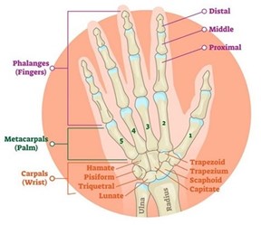
Correct Answer is C
Explanation
The bones that articulate with the carpals are the distal end of the radius and the distal end of the ulna. These two bones form the wrist joint with the carpals. Therefore, if you break the distal end of both the ulna and radius, it will result in a wrist fracture. This type of injury is also known as a distal radius and ulna fracture.
Correct Answer is A
Explanation
DNA is a double-stranded macromolecule made up of nucleotides that carry genetic information. The sequence of nucleotides in DNA determines the genetic code, which controls the development and function of living organisms. Chromosomes are structures made up of DNA and proteins that carry genes. Nucleotides are the building blocks of DNA and RNA. RNA, on the other hand, is a single-stranded macromolecule that plays a key role in protein synthesis by carrying genetic information from DNA to ribosomes.
Whether you are a student looking to ace your exams or a practicing nurse seeking to enhance your expertise , our nursing education contents will empower you with the confidence and competence to make a difference in the lives of patients and become a respected leader in the healthcare field.
Visit Naxlex, invest in your future and unlock endless possibilities with our unparalleled nursing education contents today